Introduction
Chromosomes are an essential issue in the field of cell biology, genetics as well as medical science. These are all areas which are all relevant to the USMLE. USMLE Chromosome Structure serve a crucial role in the transfer of genetic information that is vital for inheritance and helping the division of cells. Understanding chromosomes is vital for diagnosing genetic illnesses, knowing the basics of molecular biology, and understanding how cells replicate.
This article presents an extensive study of the structure of chromosomes and focuses on the most important elements of the process, and the importance of the clinical aspects that medical students should understand to successfully pass the USMLE.
1. What Are Chromosomes?
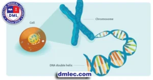
Chromosomes are a series of structures made up of DNA and proteins. In cells that are eukaryotic (like the human cell) the chromosomes reside within the nucleus. In this, they are tightly packed to protect the genetic data. Humans have 23 pairs of chromosomes (46 in total) each of which is passed down through each parent. The chromosomes contain genes, the basic components of heredity, which determine our genetic characteristics.
Prokaryotes (such like bacteria) the chromosomes of all species are circular, which makes them less complicated in arrangement. In eukaryotes, however their chromosomes are linear and joined to histone proteins, forming more complex structures.
2. The Components of Chromosome Structure
Understanding the components of chromosomes is vital to comprehend the way genetic information is stored, protected as well as transferable. Here are the most crucial components:
- DNA (Deoxyribonucleic Acid) DNA is a chemical used to encode genetic information in a double-helix structure. Every DNA molecule in the chromosome is made up of nucleotides that are sequenced. They serve as a source of instructions to create and maintain the living organism.
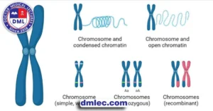
- Histones DNA is encased in proteins, referred to as histones. They aid in condensing and organizing it inside cells. The coiling of histones creates the atomic structure known as Chromin that expands further into chromosomes, which develop during cell division.
- Chromatin The fusion between DNA and histone proteins is called Chromatin. Chromatin can be found in two forms: Euchromatin, which is compact and easily accessible for transcription. Another form is heterochromatin which is packed tightly and is generally inactive.
- Nucleosome: The primary component of the chromatin. It is composed of DNA strands that are bonded to the central the core made of the histone protein. This arrangement aids in encapsulating DNA into the tiny void within the nucleus.

3. Chromosome Structure During Cell Division
In cell division, cells’ their chromosomes shrink. They can be observed under a microscope, revealing their distinct X-shaped structure. This is vital in studying the biological basis of cells genetics, as well as other USMLE related issues. The most significant structures of chromosomes are:
- Centromere is located near the middle of each chromosome. The centromere connects two sister chromatids (identical two-thirds of the entire of the chromosome ).It is essential for chromosome alignment and separation during Cell division.
- Sister Chromatids Each chromosome is split into two identical copies, called Sister Chromatids. The chromatids are joined by the centromere after which they are separated into daughter cells when they undergo mitosis.
- Telomeres They are protective caps that are located at the end of chromosomes that prevent genetic material from degrading or becoming removed during the process of cell division. Telomeres shrink when a division takes place and is related to cell death and aging.
Knowing the structure and function of such structures is crucial for medical students since anomalies or mutations in these regions could lead to cancers or genetic disorders.
4. Chromosome Banding Patterns and Identification
Chromosomes are stained with methods such as G-banding that shows both light and dark bands along the length of one’s chromosome. These bands are distinct to each chromosome. They allow scientists to identify the chromosomes which are distinct from others and regions that they are located in, as well as determine genes, and detect structural flaws. G-banding is frequently used in karyotyping. This is when the chromosomal arrangement is visible and then analysed for abnormalities.
The patterns of bands are crucial in the diagnosis of genetic disorders and are also used to map of genetic genes. Each band is a distinct DNA region, which has different levels of genes helping researchers and clinicians in the diagnosis and treatment planning.
5. Common Chromosomal Abnormalities
Chromosomal abnormalities are vital in USMLE preparation since they could be the source of many genetic disorders. These abnormalities can be classified into two types:
- Numerical Abnormalities The reason is due to an absence of or additional chromosomes. Examples include:
- Trisomy Trisomy can be described as a disorder that includes the presence of an extra chromosome (47 chromosomes ) instead of the usual 46) similar to Down syndrome (Trisomy 21).
- Monosomy A condition that results from a chromosome that is missing. It could be, for example, Turner syndrome (monosomy X) where one X chromosome is missing in females.
- Structural Abnormalities The changes are made the structure of the chromosome. They could be:
- Removals The deletion of a chromosome segment which could lead to disorders that are a result on the gene that is affected.
- Duplications The repeated sequence of a chromosome segment that result in an improve quantity of certain genes.
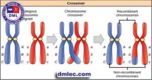
- Reversals A portion of a chromosome that has been reversed from the beginning. This may alter the function and function of the genes.
- Translocations Segments of two distinct the chromosomes can interact, which could affect the regulation of genes and cause illnesses like cancer for example.
Chromosomal abnormalities are crucial in understanding the genetic causes of disease. They are regularly examined by the USMLE to determine the impact they have upon health.
6. Clinical Relevance of Chromosome Structure in Medicine
Chromosome structures are crucial for medical diagnostics as well as the clinical setting. Techniques like KARYOTYPING and FISH (Fluorescence In-Situ Hybridization) and more advanced genetic testing allow doctors to examine and observe the chromosomes for any abnormalities that need to be detected. For example FISH employs probes that emit fluorescent light to connect specific DNA sequences to the chromosomes, which allows it to be easy to recognize and distinguish genes. This is especially beneficial for diagnosing the presence of cancer.
This test is vital to detect genetic disorders and also to determine the pattern of inheritance and offering the right genetic therapy and counseling. Chromosome analysis helps diagnose diseases early and especially those with severe or life-threatening effects.
7. Key Study Points for USMLE Chromosome Structure training
In order to be successful in the USMLE understanding the chromosome’s functions and structure is crucial. Here’s a brief overview:
- The essential components
- Learn about DNA Histones, Chromin as well as centromeres and the telomeres.
- Chromosomal anomalies
- Find out about the various kinds of abnormalities and the types of abnormalities that are common, including deletions translocations and trisomies.
- clinical methods
- Learn how to utilize Karyotyping, as well as FISH to analyze chromosomes and also diagnosing genetic disorders.
Conclusion USMLE Chromosome Structure
Chromosome structure is an intricate and crucial aspect of medical science, with direct implications in the understanding and diagnosis of genetic disorders. When they understand the structure of chromosomes, medical students gain a solid basis in genetics that will prove helpful in their entire medical job. Understanding the intricate details of chromosomes which include the components that are abnormal and diagnostic tools are vital to pass the USMLE examination in addition to other. This information helps in understanding the biological basis of genetic disorders as well as aids in the comprehension of the clinical outcome which in turn improves the treatment and care of patients.
Frequently Asked Questions USMLE Chromosome Structure
: USMLE Chromosome Structure, and Why Do They Matter for the USMLE Exam?
- Chromosomes are cell structures composed of DNA that store the genetic information needed for development, inheritance and inheritance. In terms of studying for the USMLE examinations, understanding chromosome structures is absolutely key as they form the basis of all fundamental cell biology concepts as well as genetics knowledge essential in diagnosing genetic disorders.
: The main elements of chromosome structure?
- DNA, histones (proteins that aid with coiling of DNA), Chromin (a combination of histones and DNA), nucleosomes, centromeres and Telomeres are essential components of chromosomes; each plays a vital role in storing and organizing genetic information within cells.
: The significance of centromeres and telomeres in terms of health?
- Centromeres play an essential role in keeping chromosomes aligned and aligned during cell division, while telomeres help protect both ends from degradation. Both play essential roles in maintaining cell health as well as the process of aging and replication – essential areas to study when researching genetic stability for disease or related disorders.
: How can G-banding offer when studying the chromosomes?
- G-banding uses staining techniques to reveal dark and light bands on USMLE Chromosome Structure. G-bands help identify particular chromosomes as well as regions or genes in an individual sample; G-bands also assist in pinpointing any irregularities on individual chromosomes as well as genes. This method can be especially helpful in detecting irregularities on particular chromosomes as well as for gene identification purposes.
: Which anomalies in chromosomes occur most often?
- Numerical Anomalies These conditions involve additional or missing chromosomes similar to Down syndrome (Trisomy 21) and Turner syndrome (Monosomy X).
Structure-related disorders involve changes to chromosome structures that cause duplications, deletions, translocations or inversions that alter their physical composition resulting in genetic diseases such as Duchenne muscular dystrophy (DMD).